Lensfree Angiogenesis Analysis - 6ème Congrès de la Société Française d’Angiogenèse.
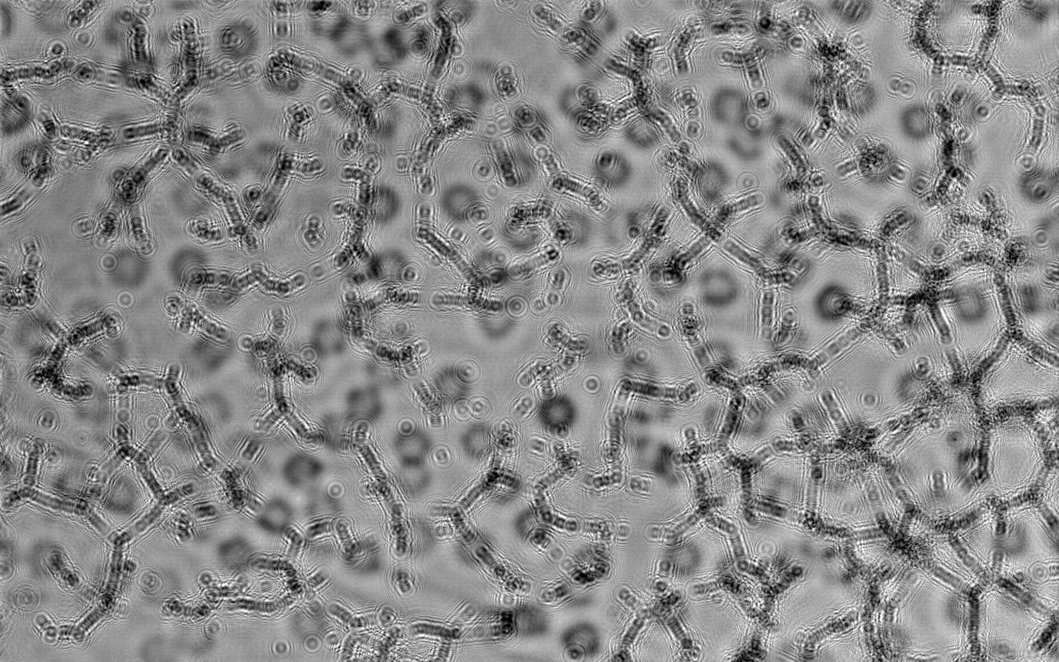
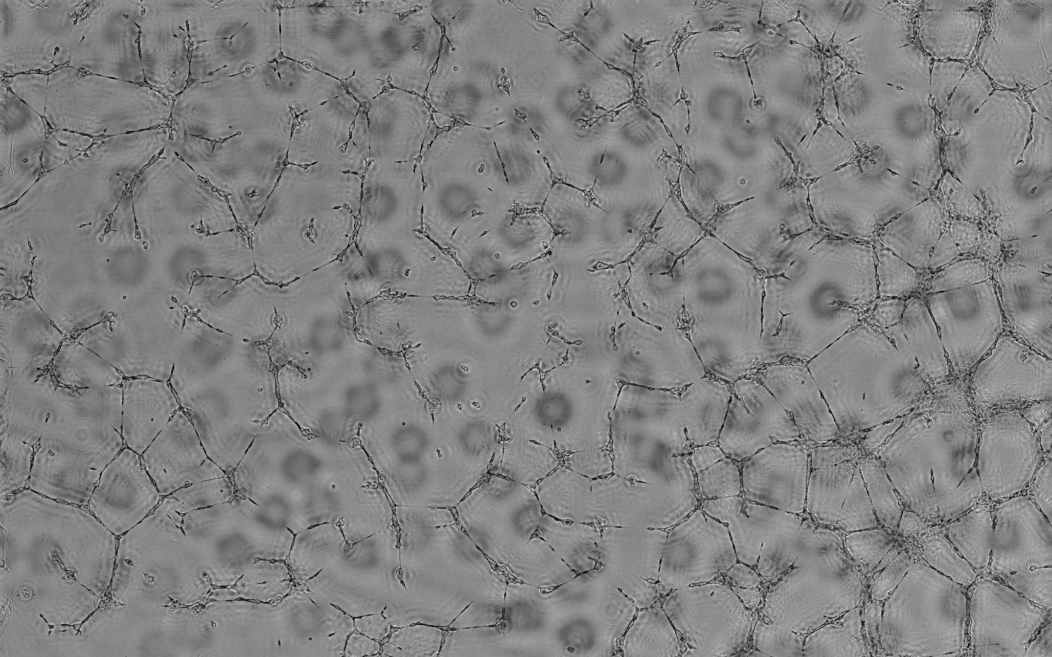
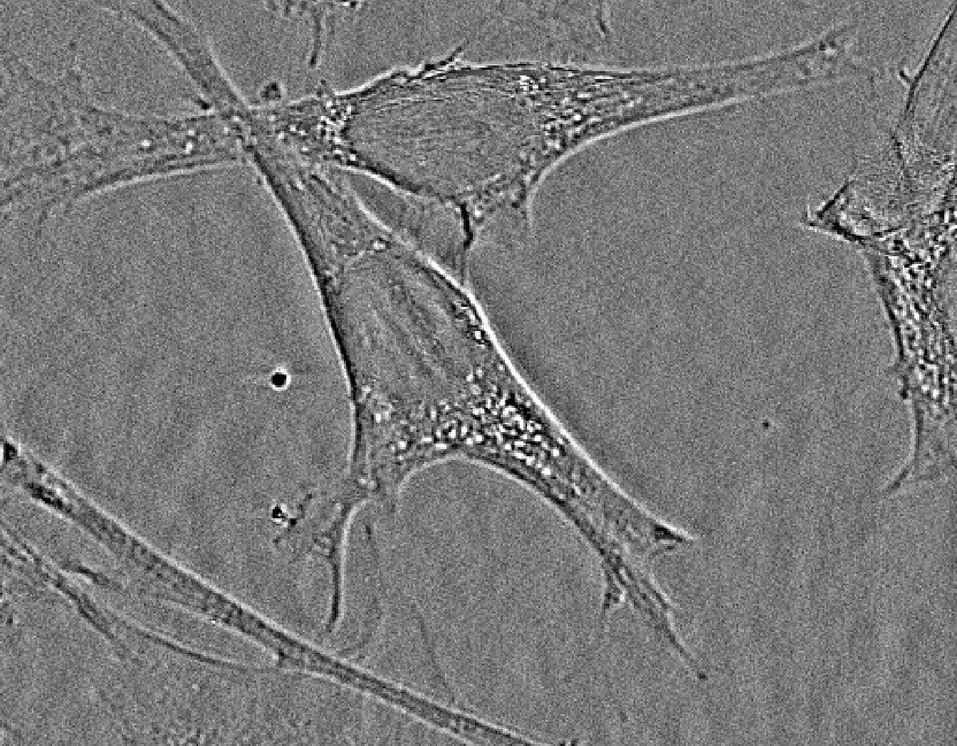
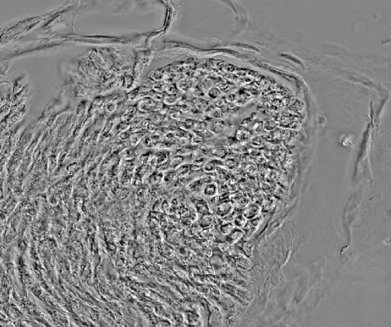
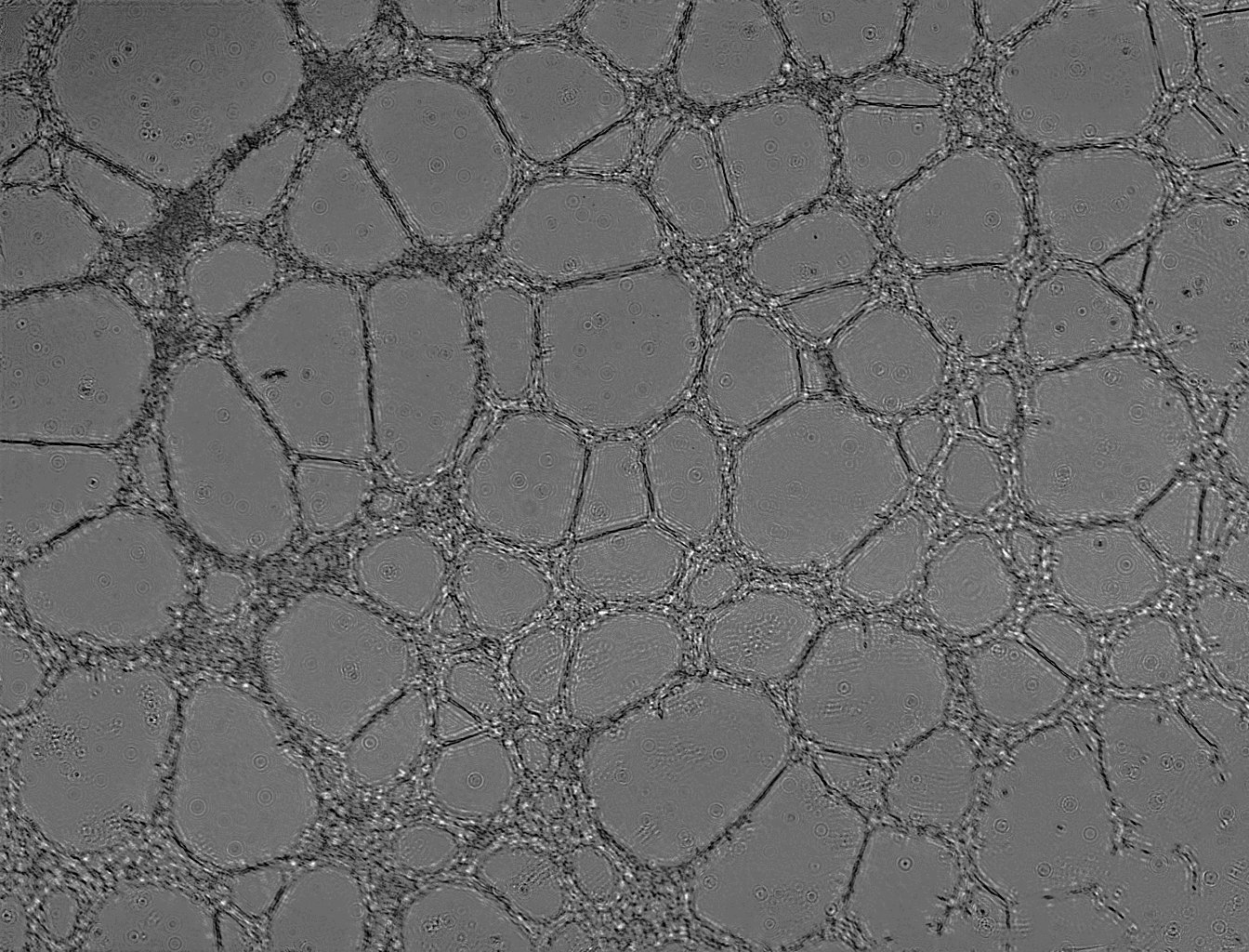
- Below: time-lapse of HUVEC culture:
Network formation with HUVEC cells observed by means of lensfree video microscopy over 47 h. The cells are cultured on 75 µl of liquid Matrigel. 5.104 HUVEC cells were then seeded on Matrigel (BD Biosciences) and incubated at 37°C. Experiment author : Boudewijn van der Sanden, PhD
Clinatec, Platform Intravital Microscopy, France Life Imaging.

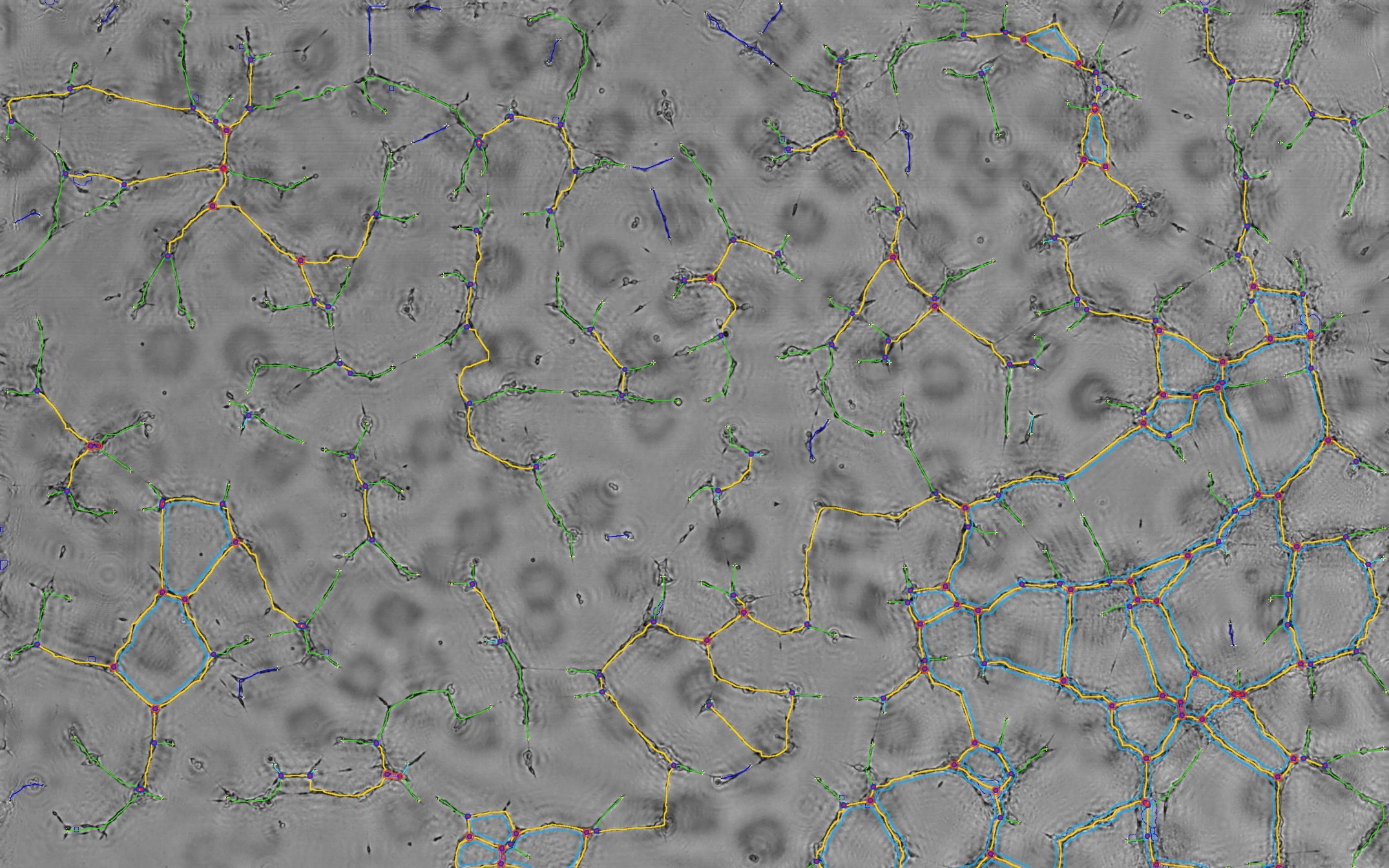
Endothelial cell network detection
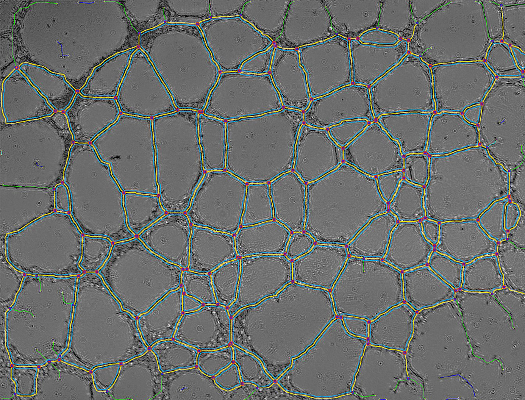
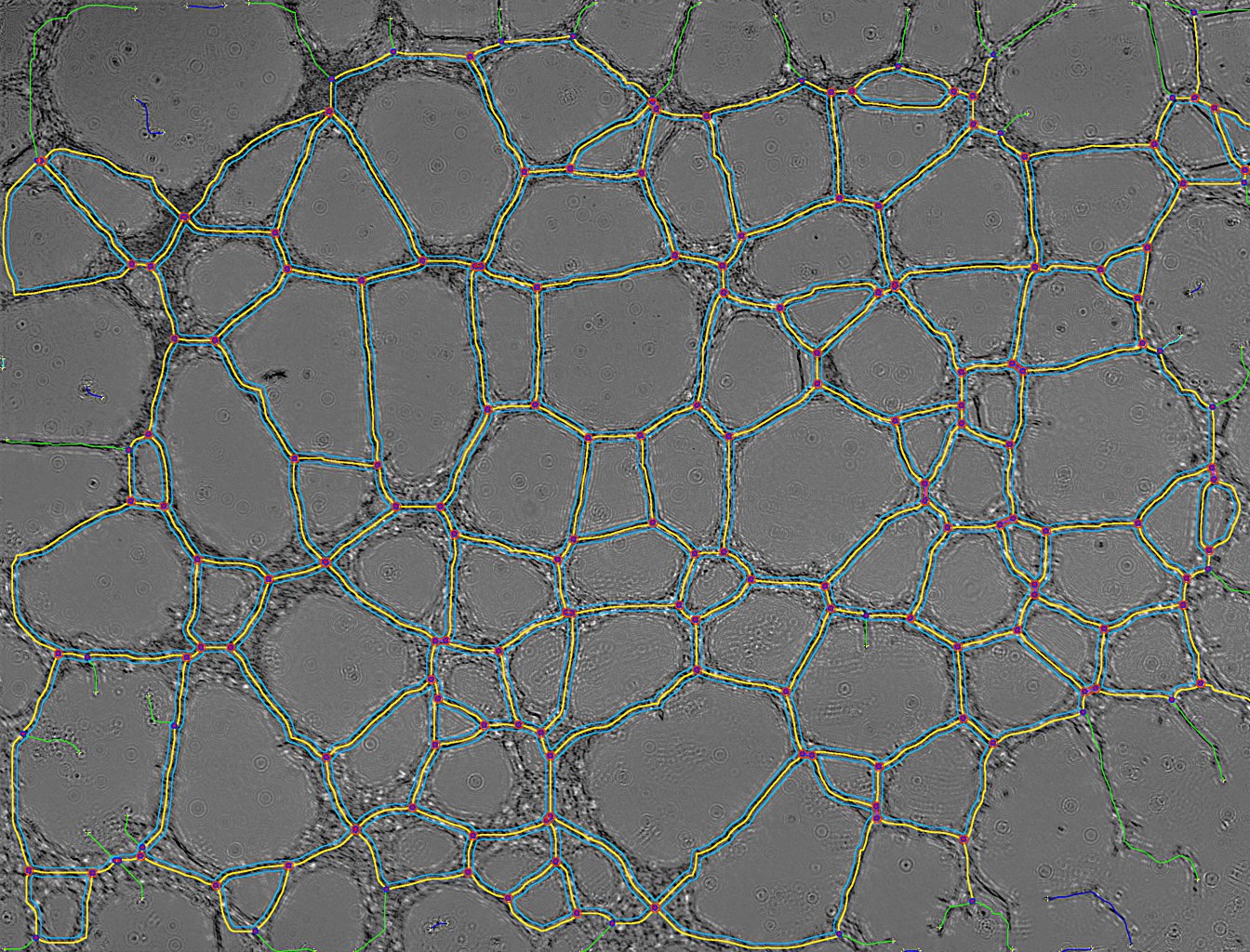
Analysis performed with a customized version of the "Angiogenesis Analyzer" ImageJ plug-in allows quantification of the meshed network into different sub-elements as shown in the slide show below :
- Below: slideshow showing analysis by the dedicated "Angiogenesis Analyzer":
segments are in yellow, branches in green, red points surrounded by blue are junctions, junctions surrounded by red are master junctions, meshes (closed structures, made by so called tube-like-structures) are in cyan and isolated structures are in blue. Orange structures are master segments (see this link for precise definition of these elements).
| prev | next |
Time-lapse analysis
- Below: movie showing a nearly 48 h analysis of HUVEC cell culture":
Lensfree Angiogenesis Analysis - hight resolution
Lensfree Angiogenesis Analysis - low resolution (smartphone, iPad...)
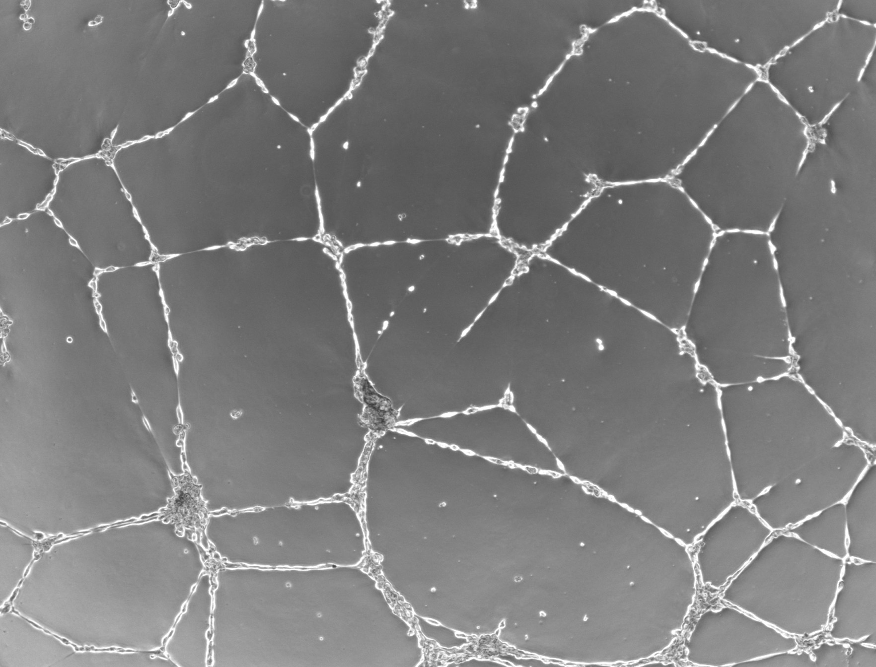
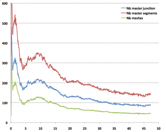
- Temporal analysis of the HUVEC network formation. The plot of the different devised metrics as a function of time clearly define three different steps for the meshed network formation: initiation, a stabilization period and then the fusion of meshes. During the first 4 hours, the network is in formation: the number of meshes increases and so do the number of segments and junctions (see above). Following this, for 6 hours the network remains stable. Towards the end, the meshes merge to form larger meshes. After 45 hours, the network presents the following architecture: total meshes area of 18 mm², with 45 meshes, with an average size of 0.4 mm² (see below).
Conclusion
Lensfree video microscope in combination with the dedicated Angiogenesis Analyzer ImageJ plug-in provides a unique mean to quantify angiogenesis and to screen ex vivo anti-angiogenic therapies. The methods is label-free, high-throughput, ease of use, working directly inside the incubator and relatively low-cost.
![]()
Collaborations

![]() Dr Cédric ALLIER. CEA-Leti - MINATEC Campus, Grenoble, France. [1]
Dr Cédric ALLIER. CEA-Leti - MINATEC Campus, Grenoble, France. [1]
![]()

![]() Geoffrey Estaban. IPRASENSE, Cap Alpha, Clapiers, France. www.iprasense.com [1]. Application for tube analysis at this link.
Geoffrey Estaban. IPRASENSE, Cap Alpha, Clapiers, France. www.iprasense.com [1]. Application for tube analysis at this link.
![]()