
Publications:
In a new publication entitled the « Weibel-Palade Bodies Orchestrate Pericytes During Angiogenesis » [1], we described a new algorithm for whole mouse retina vasculature analysis.
Whole mouse retina analysis by laser scanning microscopy:
We described in a previous work [2], an automatic analysis of P5 mouse retina vascular images obtained through laser scanning confocal imaging. The highly contrasting images allowed us to sucessfully perform these analysis (Samarelli et al. 2014).
The images obtained thanks to isolectin B4 FITC-conjugated staining, were analyzed by using a customized version of the Angiogenesis Analyzer demonstrator, (Method section, and Figure).
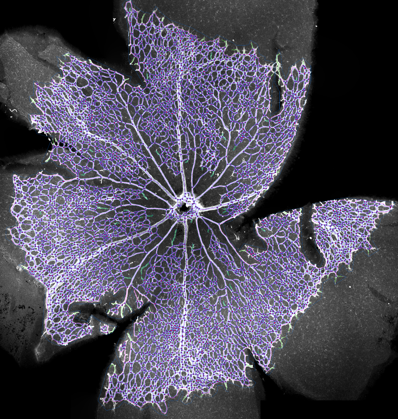
Experiment and image acquisition author: Dr. Anna Valeria Samarelli;
image analysis: Gilles Carpentier.
![]()
Whole mouse retina analysis by standard fluorescent microscopy:
In the new publication (Cossutta et al. 2019), we described an optimized segmentation method, allowing analysis of whole P5 mouse retina, using the same labelling, but from images obtained through an Orca R2 Hamamatsu CCD camera, instead of laser scanning microscopy. The algorithm wad customized to be compatible with the lower signal/noise ratio in abscence of laser illumination, and of confocal imaging. As for LSM acquisitions, images were assembled to build full retina pictures by using the MosaicJ plugin. Article and related pdf method.

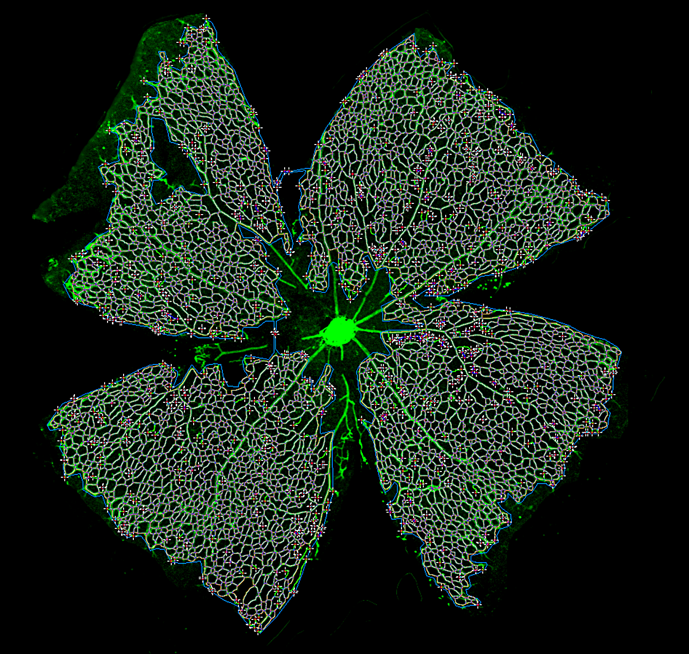
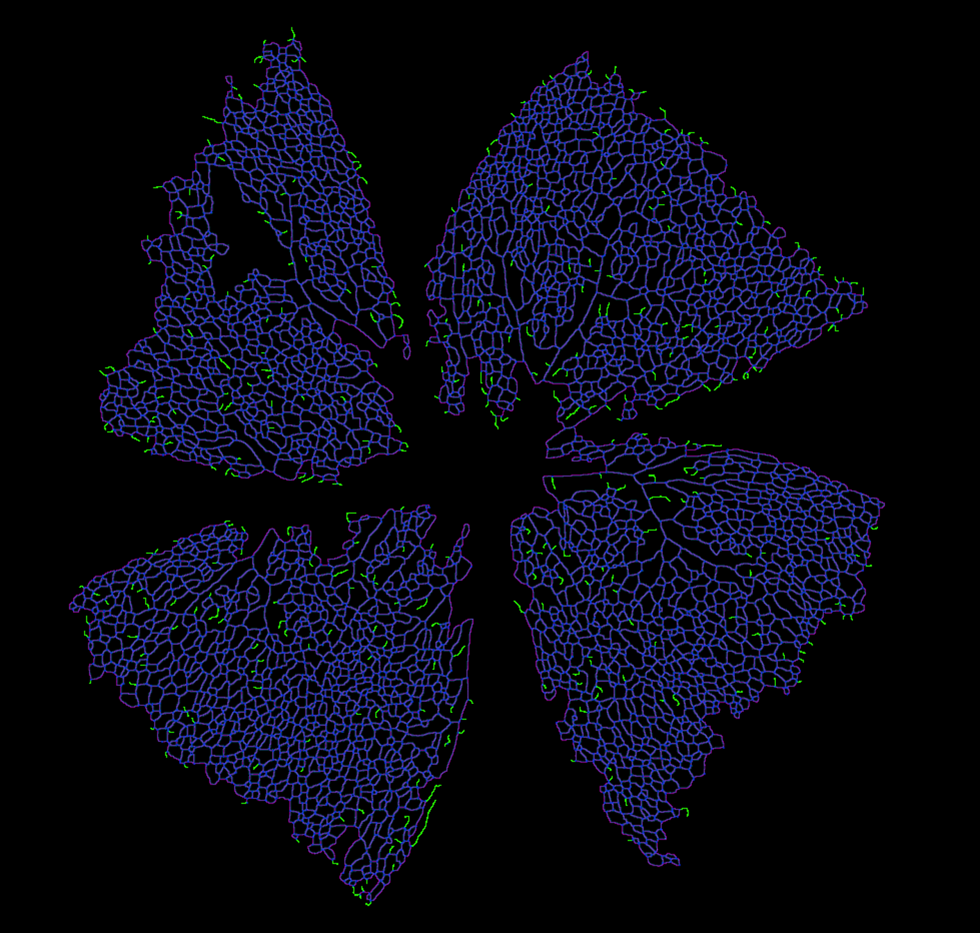


image analysis : Gilles Carpentier
![]()


