
Table of contents
- Slide show, p1
- References, p2


Slide show:
![]()


![]()


 |
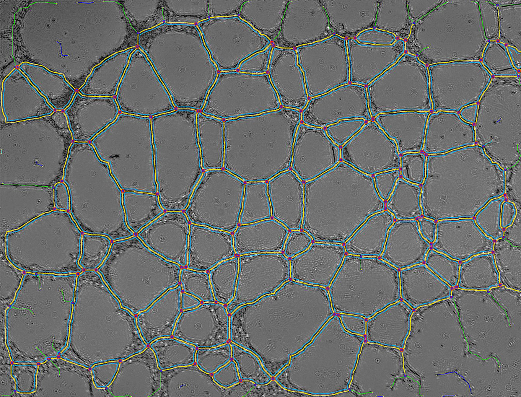
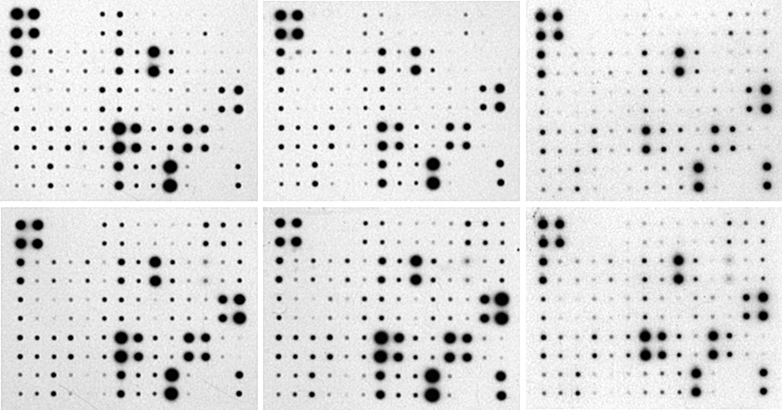
ImageJ (http://rsb.info.nih.gov/ij/) is a public domain Java image processing program inspired by NIH Image for the Macintosh. It runs, either as an online applet or as a downloadable application, on any computer with a Java 1.1 or later virtual machine. Downloadable distributions are available for Windows, Mac OS, Mac OS X and Linux. The author, Wayne Rasband (wayne@codon.nih.gov), is at the Research Services Branch, National Institute of Mental Health, Bethesda, Maryland, USA. |
 |
Gilles Carpentier, Faculte
des Sciences et Technologie, Universite Paris Est Creteil Val-de-Marne, France. Special thanks to Alessandra Albano for the English correction of these sites. |
 |
| Computer
Data Acquisition for Biochemistry Practice Works |
 Image.Bio.Methods@free.fr |
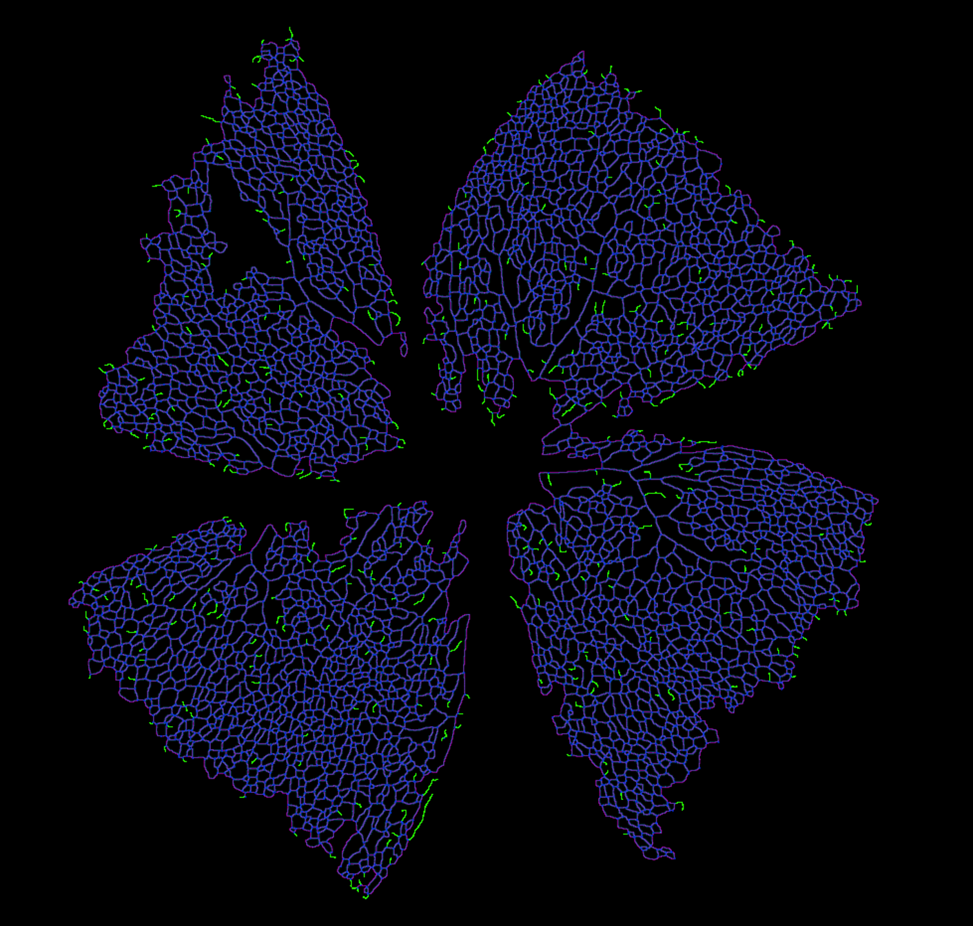
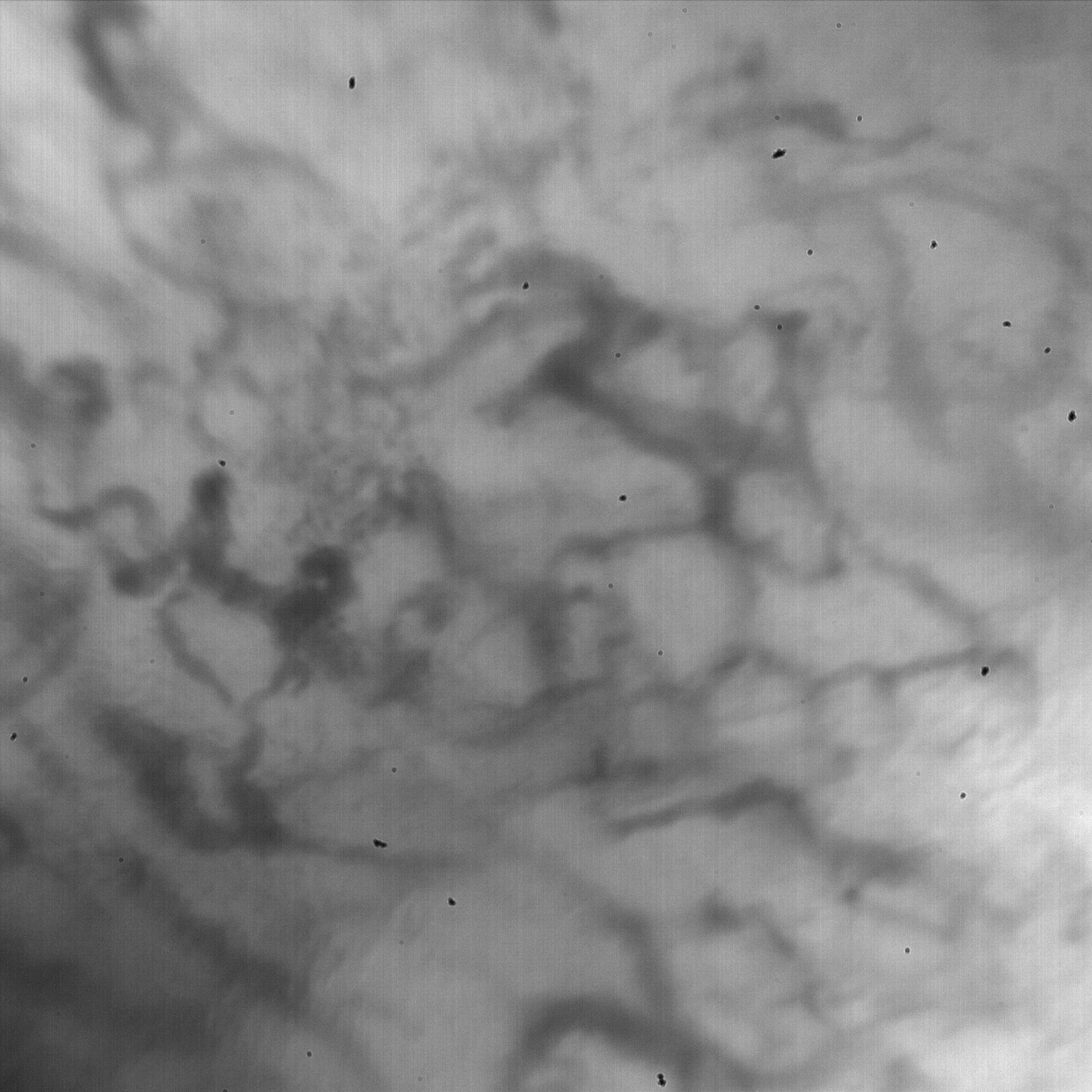
Image analysis tools in
biology and biochemistry using ImageJ |