Gilles Carpentier Research Web Site: Computer Image Analysis
Image analysis tools in biology and biochemistry using ImageJ

Home > Angiogenesis Analysis - TERMIS European Chapter Meeting 2017

Angiogenesis Analysis - TERMIS European Chapter Meeting 2017

Monday 22 May 2017, by Gilles Carpentier

Comparison of tissue-specific microvascular endothelial cells to HUVECs for skeletal muscle tissue-engineering

D. Gholobova, L. Desender, G. Carpentier, M. Gérard, L. Thorrez.

Angiogenesis Analyzer is widely used to measure in vitro angiogenesis, as shown in this non exhaustive list of reference papers. A collaboration in the field of tissue engineering of muscle has been initiated 3 years ago with Dacha Gholobova and Lieselot Decroix from the lab of Lieven Thorrez [1], [2]. Some new customizations of the Angiogenesis Analyzer lead to analyze with the same principle, endothelialization of bio-artificial skeletal muscles. The new work in this domain was exhibited at Davos in June 2017, at the TERMIS European Chapter Meeting 2017. Here after the introduction of the abstract :
PNG - 61.1 kb
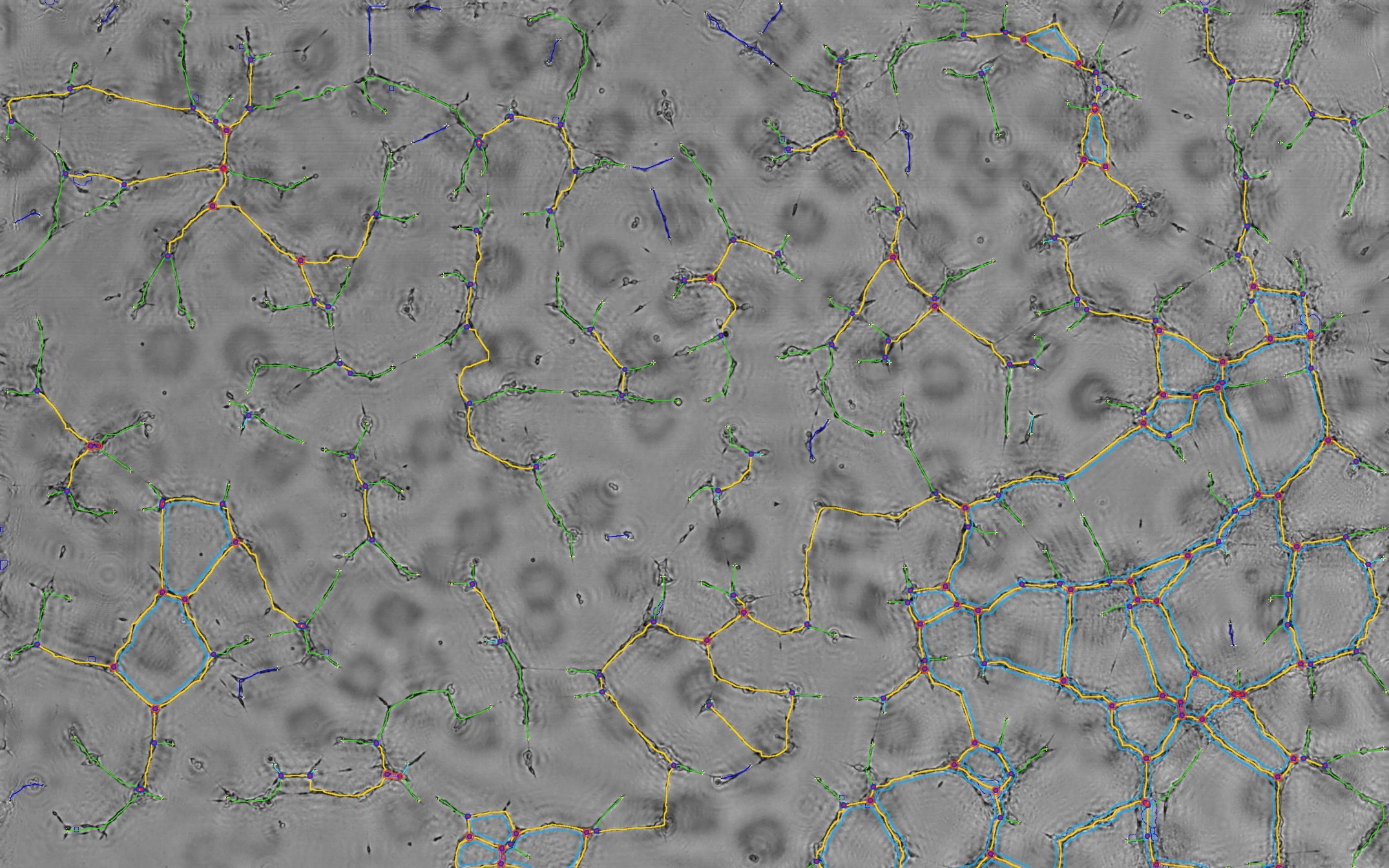
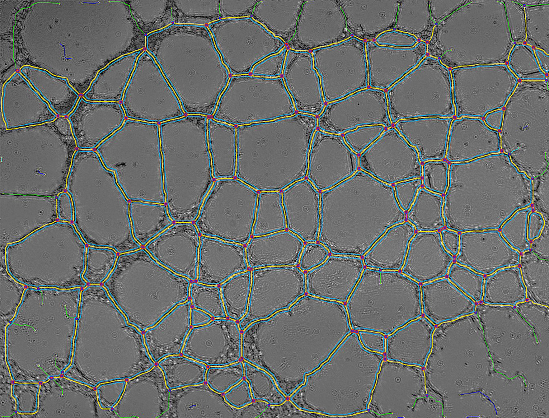
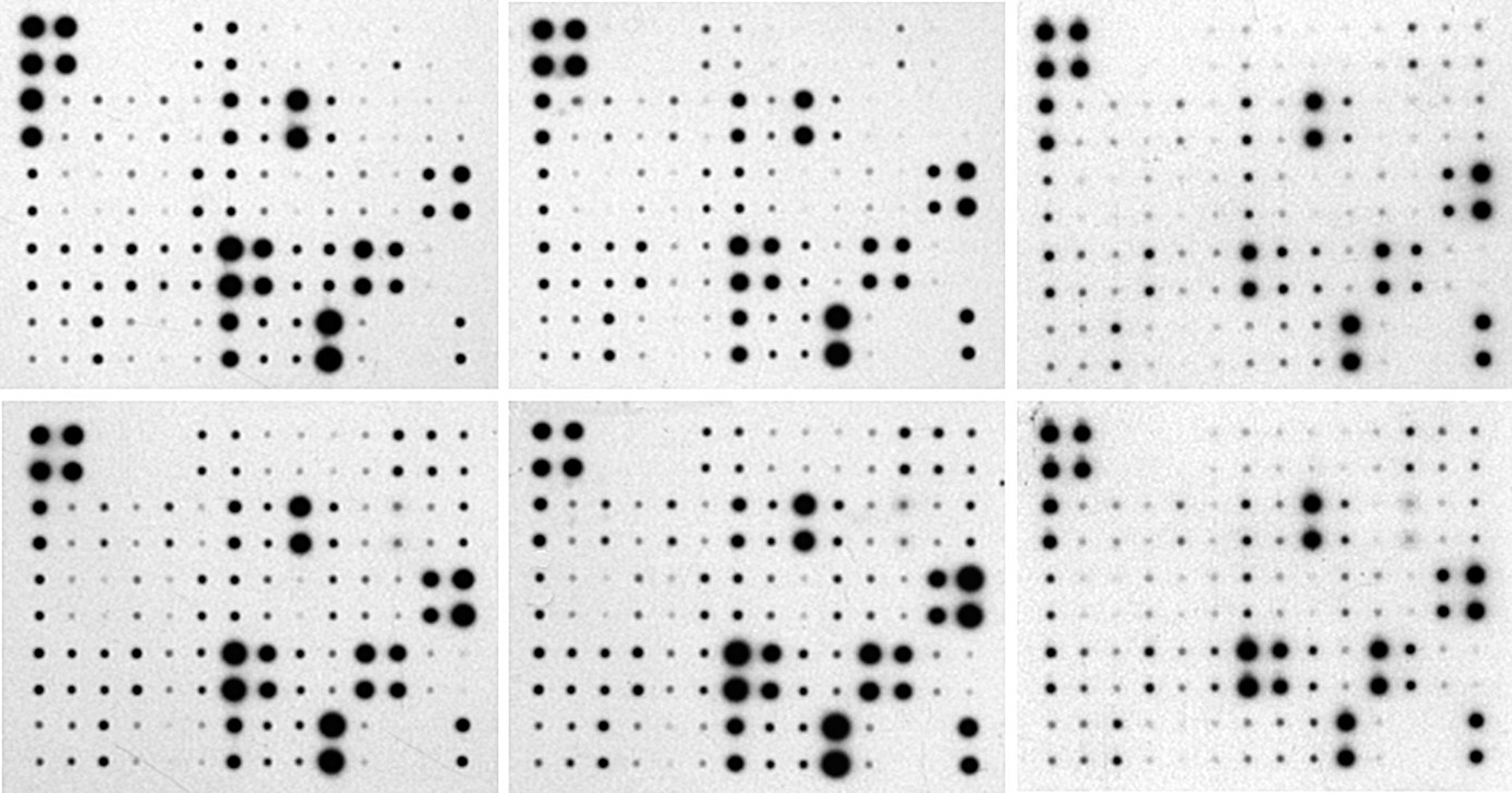
Modeled map of in-vitro endothelialization network after analysis by the customized Angiogenesis Analyzer

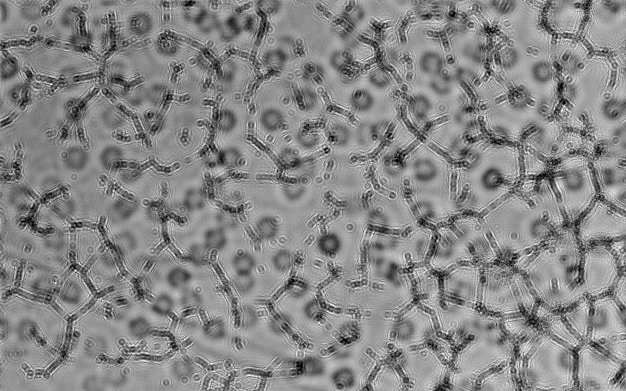
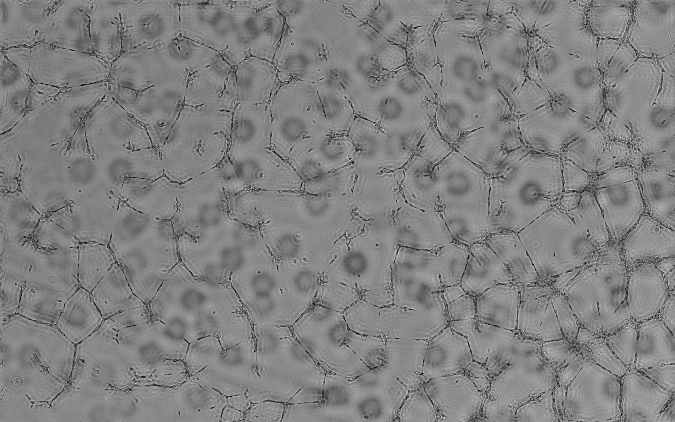
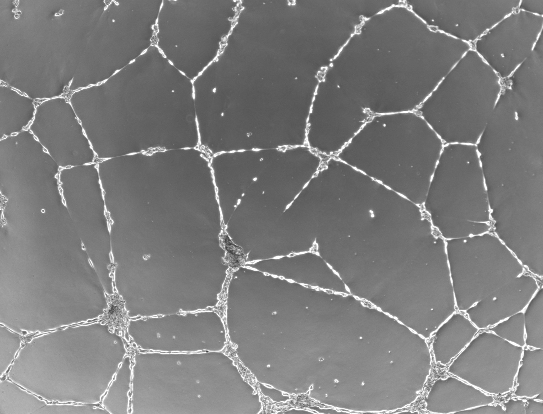
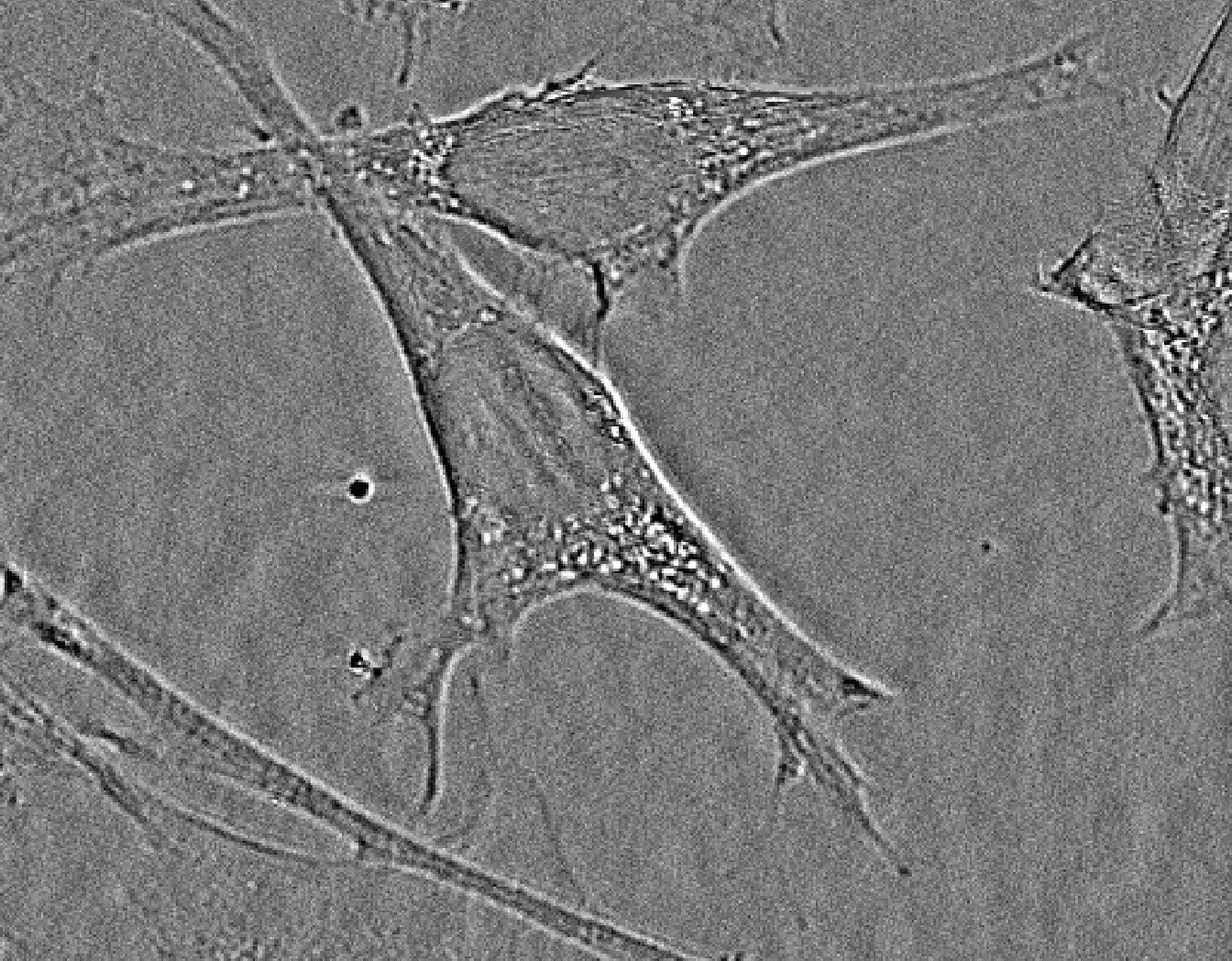
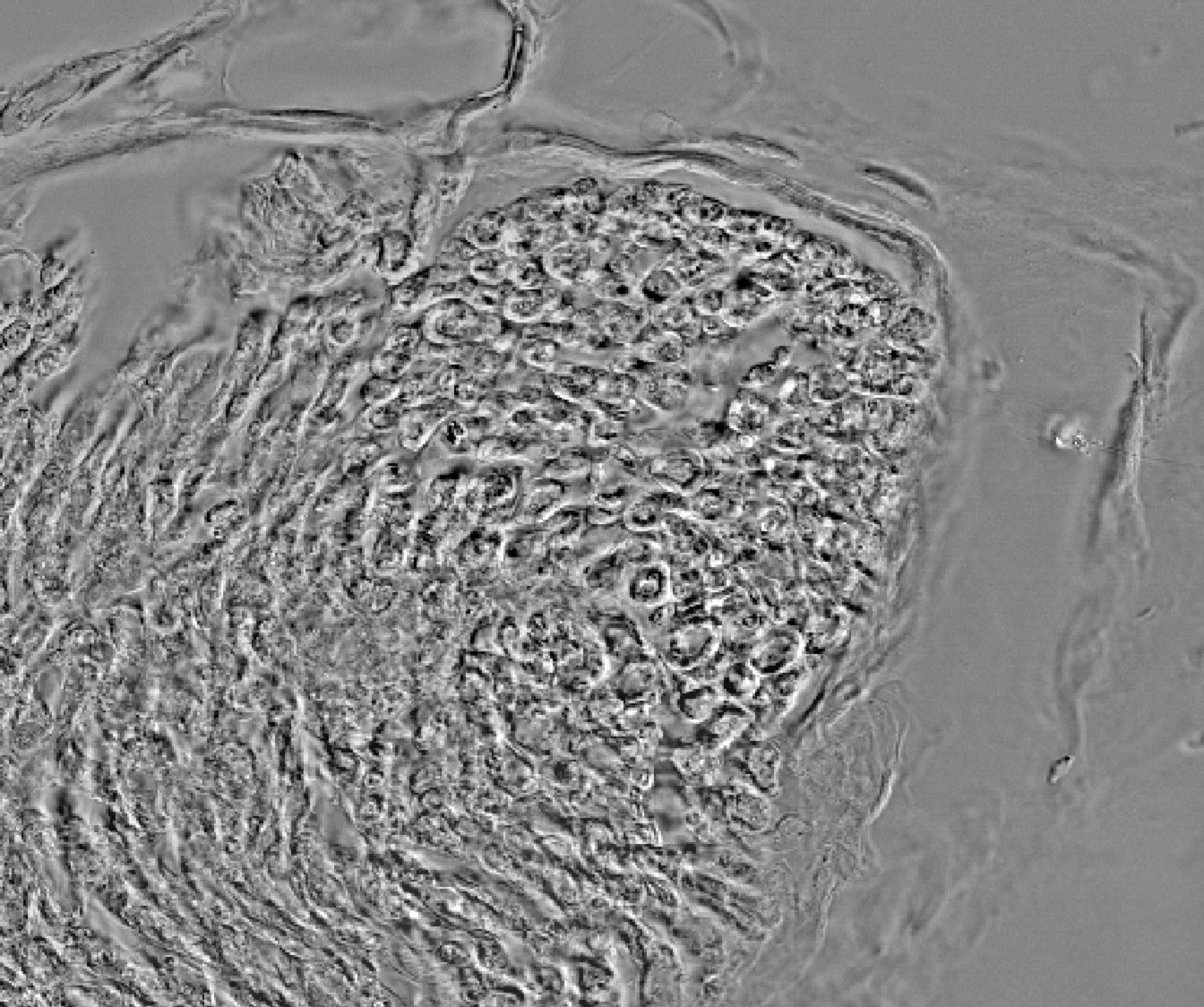
The size of in vitro engineered skeletal muscle tissue is currently still limited due to the lack of a vascular network in vitro. Different approaches for inducing vascularization in vitro have been described, but all have shortcomings. In nearly all studies, human umbilical vein endothelial cells (HUVECs) are used to develop vascular networks. HUVECs are well characterized, easy to isolate and are known for their capacity of vascular network formation in vitro. But in vivo, new blood vessel formation, angiogenesis, is mainly established by microvascular endothelial cells (MVEC) located in the smallest blood vessels, the capillaries. MVECs differ in morphologies and properties depending on the tissue. Since our interest is developing human bio-artificial skeletal muscles (BAMs), we evaluated skeletal muscle derived MVEC cells (SkMVECs) for their vasculogenic potential in our model.

Footnotes

[1Tissue Engineering Lab, Department of Development and Regeneration, KU Leuven campus Kulak, E. Sabbelaan 53, 8500 Kortrijk, Belgium - lieven.thorrez@kuleuven.be

[2Gholobova D, Decroix L, Van Muylder V, Desender L, Gerard M, Carpentier G, Vandenburgh H, Thorrez L. Endothelial Network Formation Within Human Tissue-Engineered Skeletal Muscle. Tissue Eng Part A. 2015 Sep 1. Abstract

logoij ImageJ (http://rsb.info.nih.gov/ij/) is a public domain Java image processing program inspired by NIH Image for the Macintosh. It runs, either as an online applet or as a downloadable application, on any computer with a Java 1.1 or later virtual machine. Downloadable distributions are available for Windows, Mac OS, Mac OS X and Linux. The author, Wayne Rasband (wayne@codon.nih.gov), is at the Research Services Branch, National Institute of Mental Health, Bethesda, Maryland, USA.
Gilles Carpentier, Faculte des Sciences et Technologie,
Universite Paris Est Creteil Val-de-Marne, France.

Special thanks to Alessandra Albano for the English correction of these sites.
Computer Data Acquisition
for Biochemistry Practice Works

Image.Bio.Methods@free.fr
Image analysis tools in biology
and biochemistry using ImageJ